Thursday, February 21, 2013
Tuesday, February 19, 2013
Suboccipitals: Cause of Headaches
Suboccipital Headaches: A Common Cause of Headaches
The suboccipital muscles are located at the base of your skull. Their main action is extension of your head on your neck. These muscles allow you to look up.
Many people present with a forward head posture. In this posture, one may present with rounded shoulders and a slumped upper back. The ears are not in line with the shoulders, because the head is falling forward. In order to see straight ahead, you must extend your head on your neck. This action demands the suboccipitals to contract for an extended time, which leads to muscle shortening and trigger points. This muscle tightness and trigger points may apply pressure to the occipital nerves. You can see the location of the suboccipital nerves in the pictures above. The orientation of the nerves are consistent with the location of a large percentage of headaches, especially headaches that radiate. You may even feel headache-like pain in your eyes, forehead or temple regions.
So the next time you feel this type of headache, check out your posture in a mirror. It may be the contributing factor.
Friday, February 8, 2013
The Pivot Disc
The Pivot Disc
Due to leg weakness (Knee/Hip), it may be very difficult for a
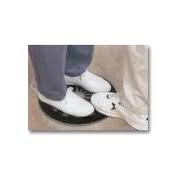
trainer, therapist or caregiver to safely transfer their client/patient from one surface to another. Their client may be able to bear weight on their legs, but may not be able to advance either foot in order to take steps. This makes transferring from the bed to a chair or vice versa very difficult. The Pivot Disc is a great tool that can be used in this situation to minimize the risk of injuring your client or yourself during functional transfers.
 |
| Pivot Disc |
 |
| Place client's feet on outer edge of disc with enough room between the feet for your foot |
 |
| Upon standing your client, use your foot that is on the disc to control the rotation of the disc, transferring your client from one surface to another Average price of a Pivot Disc is $50-60 |
Labels:
leg weakness,
physical therapy,
rehabilitation,
safety,
tools,
transfers
Monday, February 4, 2013
The VMO
 |
| Located at the inside portion of your knee |
The VMO and The Knee
The Vastus Medialis (VMO) Muscle is one of four of the the QUADriceps Muscles. The VMO is the "tear drop" shaped portion which provides a medial pull on the patella upon knee extension (tightening the thigh muscles). Upon injury, surgery, or any type of inflammation/edema to the knee joint, the VMO "shuts down." It does not take a great deal of inflammation for this to occur. Looking at the pictures below, one can see the size difference between the VMO and the Vastus Lateralis (VL). Without the VMO pulling the knee cap medially, the VL's overbearing lateral pull may lead to other knee joint problems such as Patello-Femoral Pain Syndrome. To "turn on" the VMO, many techniques and exercises are available. Neuromuscular Electric Stimulation (NMES) may be applied to the VMO and Rectus Femoris to assist muscle contraction. Tapping the VMO while performing quad exercises may also help. Check out www.kbfitnessonline.com for specific exercises that will help correct this problem
 |
| The VMO is also referred to as the "Tear Drop" shaped muscle |
 |
| Vastus Lateralis is a much larger muscle than the VMO |
 |
| Here's is picture of the VMO and VL with Quad Contraction |
Subscribe to:
Comments (Atom)


